
By
Dr Digish Shah
Senior Consultant Nuclear Medicine
The diagnosis and staging of cancer have significantly evolved due to advancements in imaging technology. Among these, positron emission tomography (PET) has emerged as a powerful tool in modern oncology.
When combined with CT or MRI, PET imaging provides both functional and anatomical insights, helping doctors make more accurate decisions.
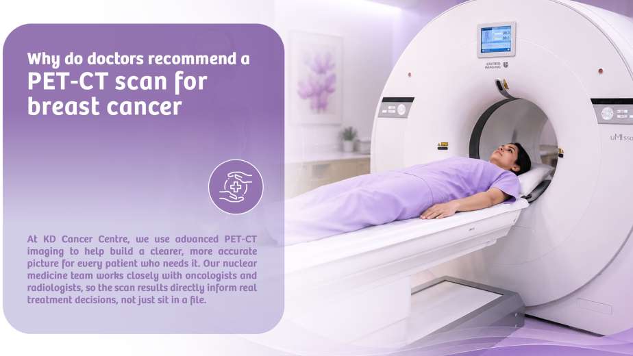
In this blog, we will explore how PET imaging improves cancer staging, along with its benefits, limitations, and the growing role of nuclear medicine in cancer diagnosis. It is particularly relevant for patients seeking advanced care at a nuclear imaging centre in Ahmedabad.
What is PET Imaging in Cancer Diagnosis?
PET imaging is a type of nuclear medicine scan that uses a small amount of radioactive tracer. Most commonly FDG (fluorodeoxyglucose) to detect metabolic activity in the body. Cancer cells tend to have higher metabolic rates than normal cells, which makes them appear as bright spots on PET scans.
Unlike traditional imaging methods that focus on structure, PET scans highlight how tissues and organs are functioning, making it particularly useful for detecting cancer early and assessing its spread.
Role of Nuclear Medicine in Cancer Diagnosis
Nuclear medicine has transformed the way cancers are detected and monitored. It enables clinicians to
- Identify cancer at an early stage
- Determine the extent of disease spread
- Evaluate treatment response
- Detect recurrence after treatment
PET imaging is one of the most advanced applications of nuclear medicine, offering unmatched sensitivity in identifying active cancer cells.
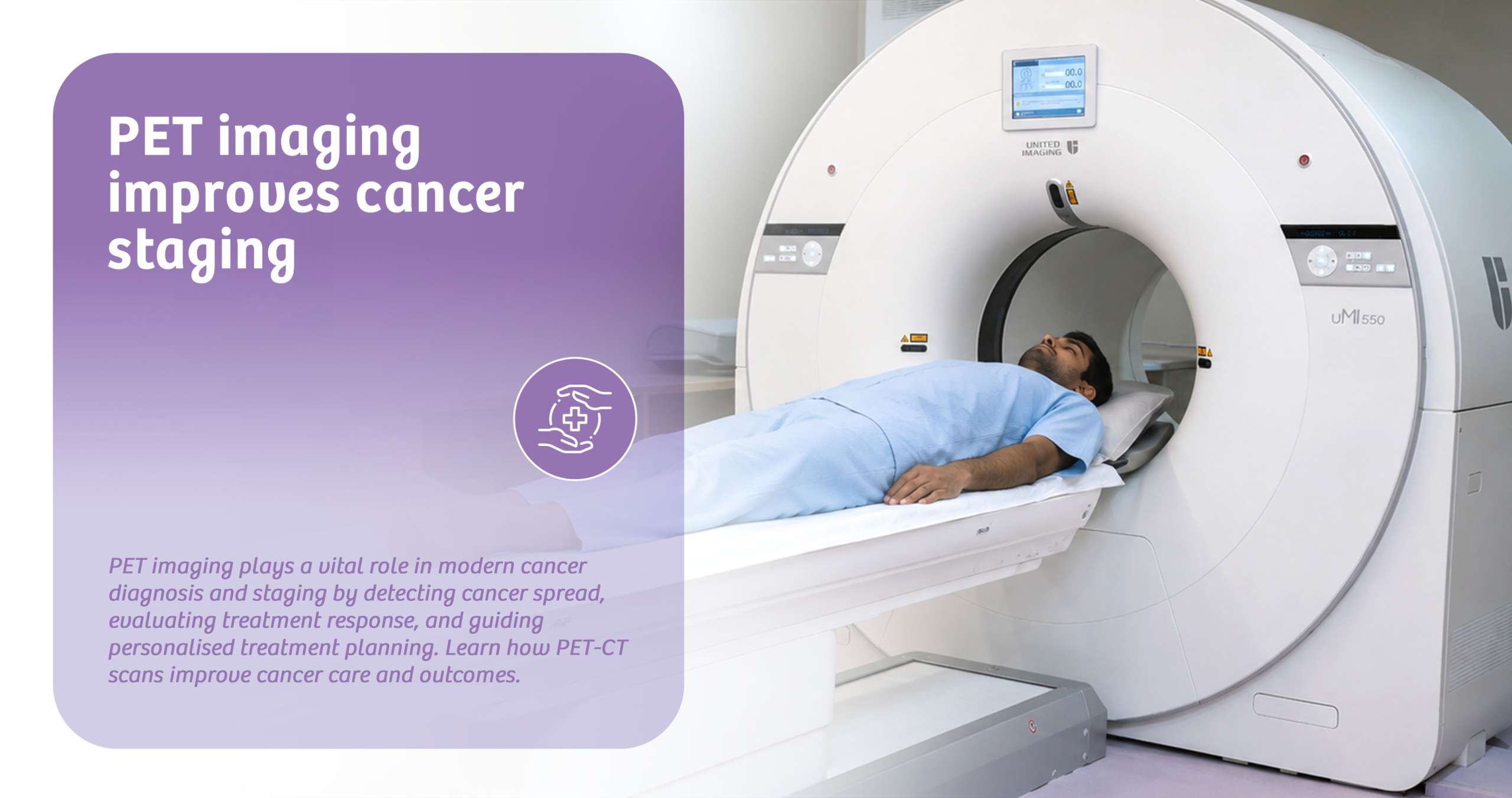
How PET imaging improves cancer staging
Cancer staging is the process of determining the extent of disease progression and how far it has spread within the body. PET imaging plays a crucial role in this process in several ways
1. Detects early spread (Metastasis)
PET scans can identify cancer spread to lymph nodes or distant organs even before structural changes become visible on CT or MRI.
2. Provides whole-body imaging
A PET scan evaluates the entire body in a single session, helping doctors assess whether cancer has spread beyond the primary site.
3. Differentiates between benign and malignant lesions
By proper analysis of scan images and based on experience, the difference between benign and malignant lesions can be determined.
4. Improves the accuracy of staging
Accurate staging is essential for selecting the right treatment plan—whether surgery, chemotherapy, radiation, or targeted therapy.
5. Helps in treatment planning
PET imaging enables oncologists to precisely locate tumours, particularly for planning radiation therapy.
Benefits of PET scan for cancer detection
- High Sensitivity and Accuracy
PET scans detect even small clusters of cancer cells due to their increased metabolic activity.
- Early Detection
PET imaging can reveal cancer before it becomes visible on conventional scans.
- Better Treatment Monitoring
Doctors can assess how well a treatment is working by observing changes in metabolic activity.
- Minimally Invasive
The procedure involves only a small injection of tracer and does not require surgery.
- Whole-body Assessment
Useful for cancers that tend to spread, such as lymphoma, lung cancer, and colorectal cancer.
When is a PET Scan Recommended?
Doctors may Advise a PET Scan in the following Situations
- To confirm or detect cancer
- For staging and assessing spread
- To evaluate treatment effectiveness
- To detect recurrence after treatment
- To guide biopsy or radiation therapy
What to Expect During a PET scan?
- A small amount of radioactive tracer is injected into the body
- You may need to wait 60-90 minutes for the tracer to distribute
- The scan itself takes about 20–40 minutes
- The procedure is painless and non-invasive
Patients should refrain from eating for a few hours before the scan and avoid strenuous activities beforehand.
Why we?
If you are considering a PET scan for cancer detection, choosing the right facility is crucial. A well-equipped nuclear imaging centre should offer
- Advanced Digital PET-CT technology
- Experienced nuclear medicine specialists
- Accurate and timely reporting
- Comprehensive cancer care support
In a nutshell
PET imaging has revolutionised cancer diagnosis and staging by providing detailed insights into how cancer behaves in the body. Its ability to detect early spread, assess treatment response, and guide clinical decisions makes it an indispensable tool in modern oncology.
While it has certain limitations, the benefits of PET scans far outweigh the drawbacks, especially when combined with CT imaging. For patients seeking accurate cancer diagnosis and staging, consulting a trusted nuclear imaging centre in Ahmedabad can make a significant difference in treatment outcomes.
FAQs
1. Is a PET scan safe for cancer detection?
Yes, PET scans are generally safe, with minimal radiation exposure.
2. How long does a PET scan take?
The entire process may take 2-3 hours, including preparation time.
3. Can a PET scan detect all types of cancer?
Most cancers can be detected, but some tumours may be missed due to differences in the biological behaviour of the disease and the radiopharmaceutical used for the scan.
4. Do I need to prepare for a PET scan?
Yes, fasting for a few hours and avoiding heavy exercise is usually recommended.